Phase-contrast & dark-field imaging
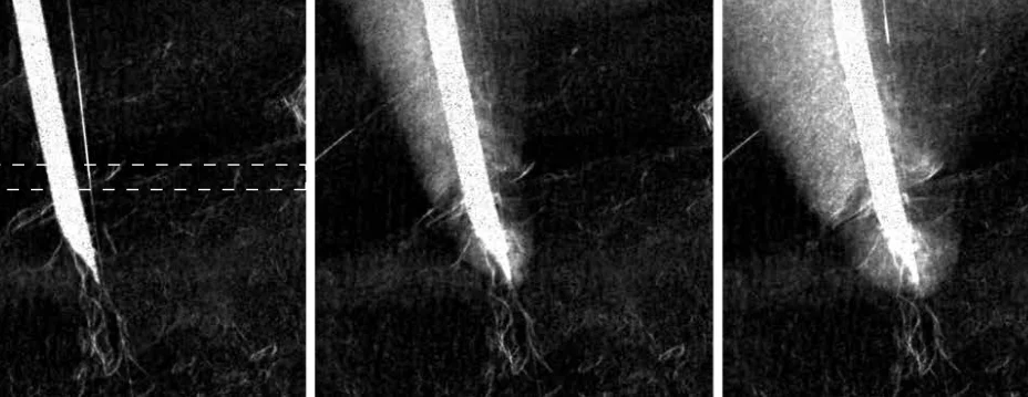
Exploiting the refractive index and small-angle scattering of X-rays for contrast channels beyond conventional attenuation, both at grating-based lab setups and at synchrotrons.
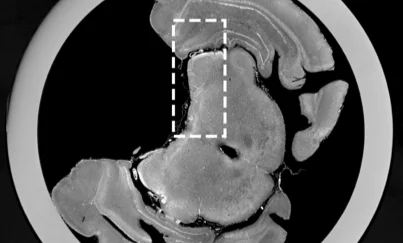
Fig. from IEEE TIP 2026 · CC-BY
Physicist · X-ray imaging researcher
I work on improving X-ray imaging methods to extract more information from medical and scientific images, using techniques that can reveal soft tissue, microscopic structure, and details that conventional scanners miss.
I’m a physicist and researcher at the Technical University of Munich, working on phase-contrast and dark-field X-ray imaging. These techniques turn what a conventional scanner throws away (the wave-like behavior of X-rays) into new contrast channels for medical and scientific imaging.
In practice this is a mix of applied physics, instrumentation, and data analysis: designing better acquisition schemes, writing data processing algorithms, and figuring out what the resulting images actually tell us. So far I’ve worked on dynamic dark-field tomography for monitoring cryoablation, high-efficiency phase microtomography at synchrotron sources, and X-ray virtual histology.
I like clear explanations and building things that actually work. I’m always open to collaborations, so if you have an interesting sample, an imaging problem, or an idea that might benefit from these methods, feel free to get in touch!
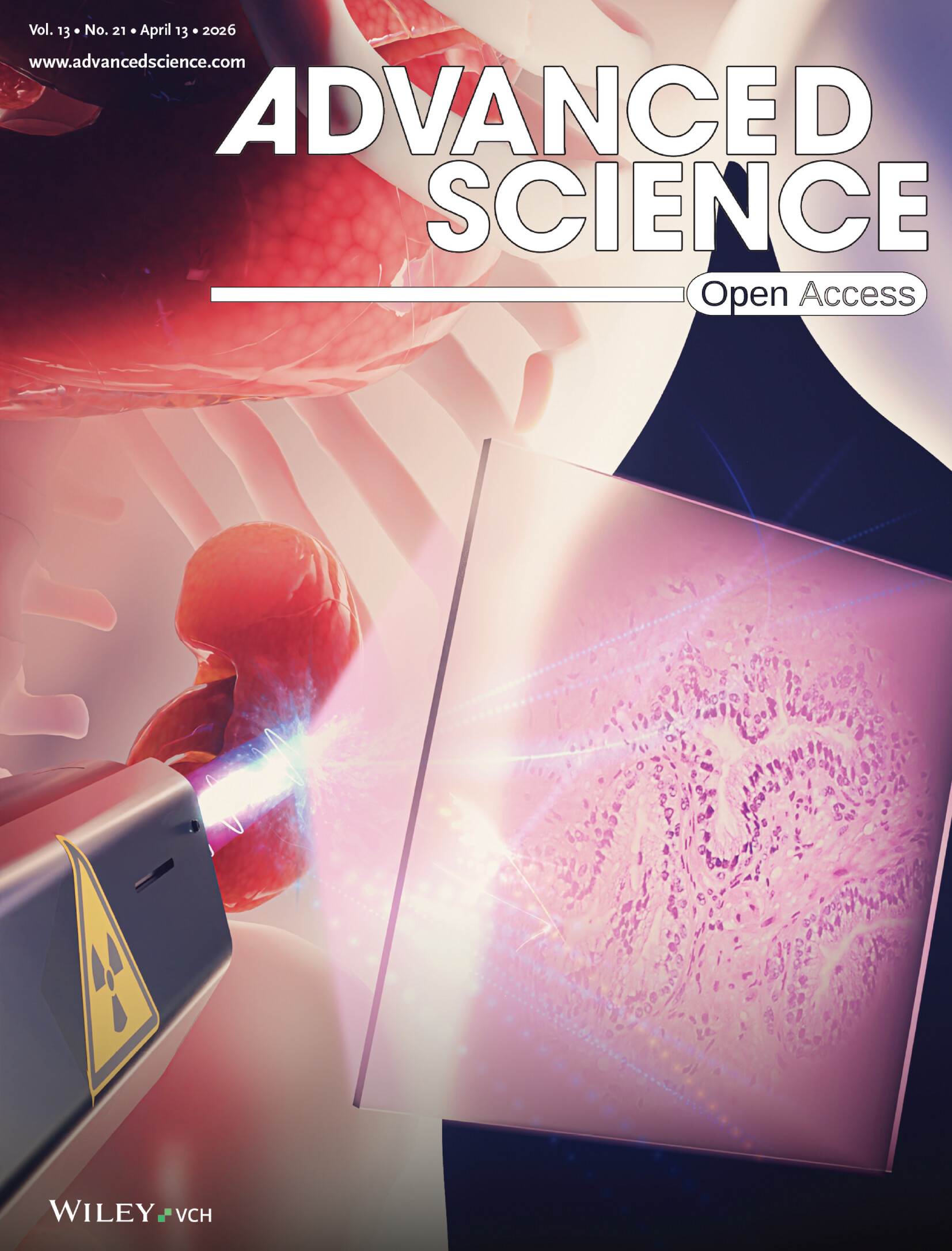
Cover article
Advanced Science 13, 21 (2026) · Article e74957
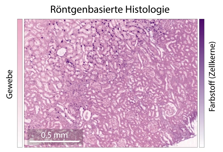
A method that generates microscopy-style histological images directly from 3D X-ray scans of excised tissue — letting researchers navigate a specimen’s full 3D volume in a familiar histological format, without destroying it in the process.
Exploiting the refractive index and small-angle scattering of X-rays for contrast channels beyond conventional attenuation, both at grating-based lab setups and at synchrotrons.
Fig. from IEEE TIP 2026 · CC-BY
Getting anatomical and tissue-level information out of 3D X-ray scans to aid biomedical research and diagnosis, without physically cutting the sample the way conventional histology requires.
Multimodal monitoring of dynamic processes, by adding phase-contrast and dark-field channels to conventional CT for enhanced contrast and quantitative information. One example: tracking tissue cryoablation as it happens.
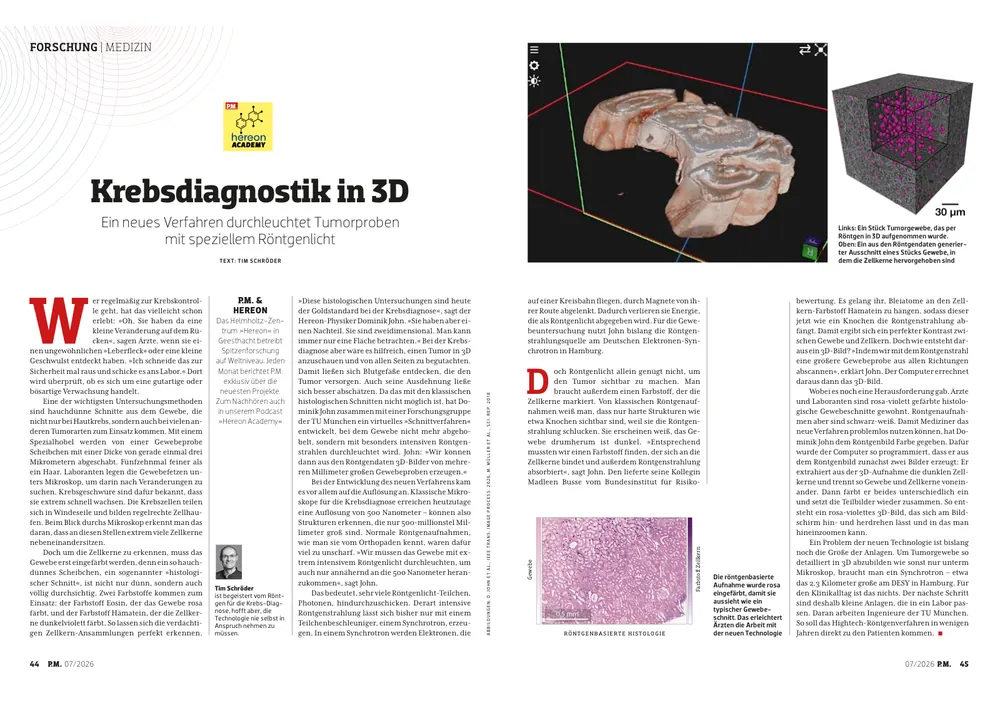
P.M. magazine · Jul 2026
Interview about my virtual histology research in Germany’s second-largest popular-science magazine. We discuss how 3D X-ray imaging of excised tissue could improve cancer diagnostics, letting doctors examine tumors without slicing them into thin sections.
TUM press release · Mar 2026
TUM feature on an advanced X-ray imaging technique that extracts more information from biological specimens while lowering radiation dose.
Hereon press release · Jan 2026
Press release on a 3D X-ray method that brings standard histology staining together with whole-specimen imaging, so researchers can see tissue structure in 3D rather than only in thin 2D sections.
FastForwardScience · 2025
Award-winning short explainer video on why medical X-rays have been monochrome for over a century and how spectral X-ray imaging is finally adding color. The technique distinguishes materials by their energy-dependent attenuation, with the goal of catching disease earlier and more reliably.
Young Scientist Award — Long Best Debut Video · FastForwardScience 2025
Photo: Simon Esser / Wissenschaft im Dialog
Peer-reviewed. Full, up-to-date list on ORCID and Google Scholar.
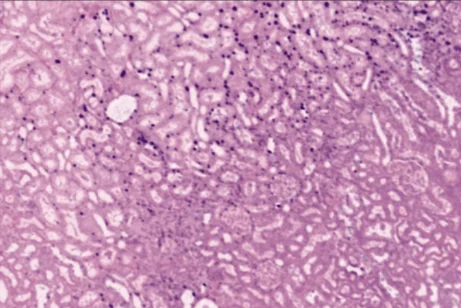
Quantitative Stain Mapping in X-Ray Virtual Histology
Advanced Science 13(21), e19783 · 2026 Hereon press
Near-perfect efficiency in X-ray phase microtomography
Optica 13, 273–283 · 2026 TUM press
Extending the field of view in modulation-based X-ray phase microtomography
IEEE Transactions on Image Processing · 2026
X-ray dark-field computed tomography for monitoring of tissue freezing
Scientific Reports 14, 5599 · 2024
Directional Dark Field for Nanoscale Full-Field Transmission X-Ray Microscopy
Light: Science & Applications · 2026
Self-supervised denoising of grating-based phase-contrast computed tomography
Scientific Reports 14, 32169 · 2024
Medical Physics 52(4), 2145–2154 · 2025
Recent developments in quantitative phase-contrast microtomography using Talbot Array Illuminators
Proc. SPIE — Developments in X-Ray Tomography XV, 13152, 1315215 · 2024
Quantitative Virtual Histology using Single-Grid X-Ray Microtomography
International Conference on X-ray and Neutron Phase Imaging with Gratings (XNPIG) · Munich, Germany · Jun 2026
Quantitative stain mapping in X-ray virtual histology
21st European Molecular Imaging Meeting (EMIM) · Ljubljana, Slovenia · Mar 2026
High-resolution phase-contrast imaging for large samples at synchrotron sources with time-varying beam profiles
International Symposium on Medical Applications of X-ray Phase-Contrast & Photon-Counting (IMXP) · Munich, Germany · Jun 2024
X-ray dark-field computed tomography for monitoring of tissue freezing
International Conference on X-ray and Neutron Phase Imaging with Gratings (XNPIG) · Shenzhen, China · Apr 2024
Recent developments in quantitative phase-contrast microtomography using Talbot Array Illuminators
SPIE Optics + Photonics · San Diego, USA · Aug 2024
Wavefront-marker phase-contrast imaging for centimeter-sized samples
15th International Conference on Synchrotron Radiation Instrumentation (SRI) · Hamburg, Germany · Aug 2024
Quantitative phase-contrast microtomography at Beamline P05 (PETRA III)
International Symposium on Medical Applications of X-ray Phase-Contrast & Photon-Counting (IMXP) · Munich, Germany · Jul 2023
Fate and effects of tattoo pigments in human and porcine skin
16th International Conference on X-Ray Microscopy (XRM) · Lund, Sweden · 2024
Essays on Medium about the surprising physics of everyday things, and the stories behind why we believe what we believe.
The best way to reach me is by email: mail [at] dominikjohn.com.
Technical University of Munich · Munich, Germany